
College of Science & Health (CSH)
Imaging and surface analysis
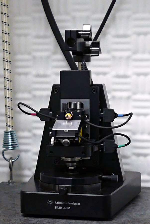
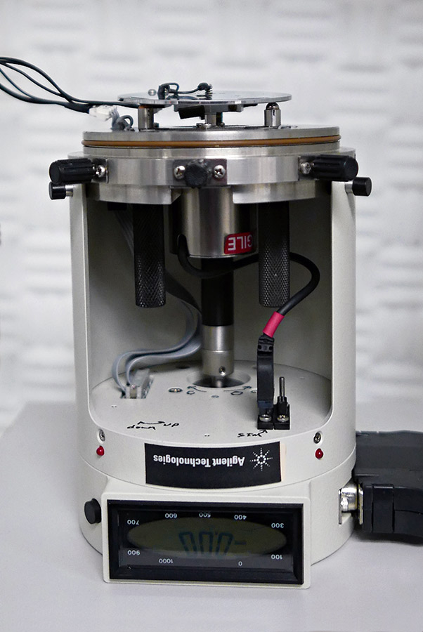
Atomic Force Microscope (AFM)
The AFM maps nanoscale topography by scanning a sharp tip back and forth across a surface. The tip is mounted on a flexible cantilever that is able to apply force to the surface, depending on the stiffness of the cantilever. It produces dimensional images with angstrom-scale variations in height and nanometer scale spatial resolution. In addition to imaging, roughness and force profiles can be obtained.
Agilent 5420
- Contact and noncontact imaging
- PicoView and PicoImage software
- Internal MAC Mode III controller
- Samples can be imaged in air, liquid, or controlled gas environments
- Variety of scanners and nosecones to enable imaging of a wide range of samples
- Operates at temperatures from -10 C to +200 C
- Scanner head approaches sample
Contact: Aric Opdahl
 Agilent-5420
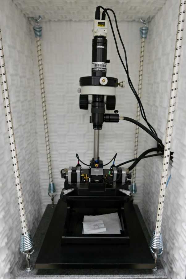
Agilent 5500
- Contact and noncontact imaging
- PicoView and PicoImage software
- External MAC Mode III controller
- Samples can be imaged in air, liquid, or controlled gas environments
- Variety of scanners and nosecones to enable imaging of a wide range of samples
- Operates at temperatures from -10 C to +200 C
- Sample approaches scanner head
Contact: Seth King
 Agilent 5500
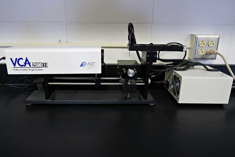
Contact Angle Goniometer
The goniometer is used to measure the surface tension/energies of liquids and solids. For example, the instrument can be used to characterize the hydrophobic or hydrophilic nature of a surface.
AST 2500
- Manual drop setting
- Can be used with a variety of liquid and solid samples
Contact: Aric Opdahl
 AST-2500
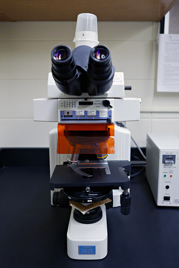
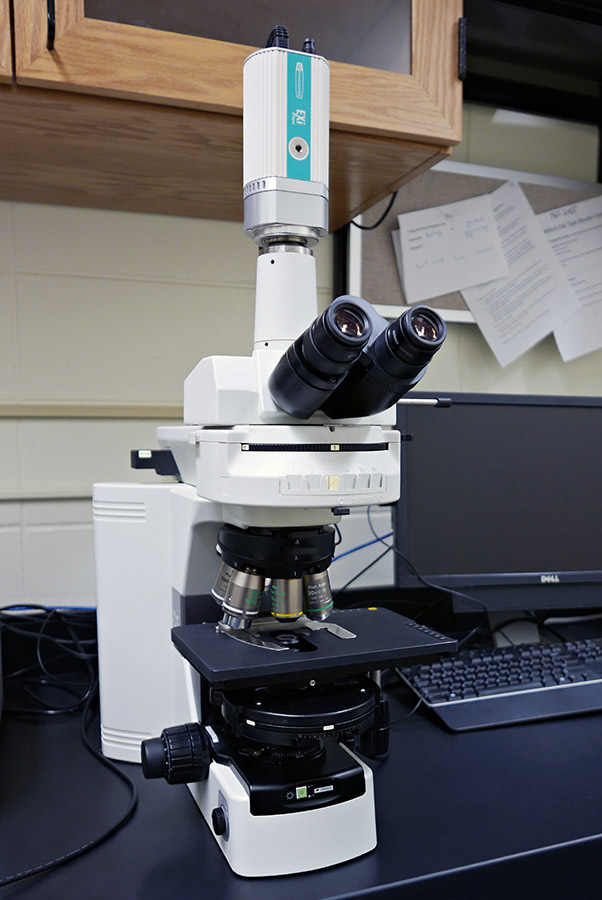
Fluorescence Microscope
This microscope is used to image fluorescent samples. Images are taken of emitted light from the sample after being hit with a beam of light at a sufficient wavelength to incite excitation. Samples that are not autofluorescent are prestained with a fluorescent dye before imaging. Commonly used for biological samples (e.g. cells, platelets).
Nikon Eclipse E600
- Upright compound microscope
- 10, 20, 40, 60, 100x objectives
- Differential interference contrast (DIC) on 40-100x
- Filters for blue, green, and red fluorescence
Contact: Tony Sanderfoot or Sarah Lantvit
 Nikon-Eclipse-E600
Nikon Eclipse 80i
- Upright compound microscope
- 2, 10, 20, 40, 60, 100x objectives
- DIC on 20-100x
- Darkfield on 10 and 40x
- Filters for blue, green, red, and far red fluorescence
Contact: Tony Sanderfoot or Sarah Lantvit
 Nikon-Eclipse-80i
Cameras
- 3 cooled CCD cameras for fluorescence and gray scale imaging
- 2 research grade color CCD cameras
- 1 high speed digital video camera
3rd Fluorescent Microscope Calendar
Laser Scanning Confocal Fluorescence Microscope
Used to image fluorescent samples. Confocal microscopy allows for scanning a sample's surface with a concentrated beam, instead of illuminating the entirety of the sample at once. This allows for increased quality of images with greater resolution over a traditional fluorescence microscope.
Leica Stellaris 5
- DMI8 inverted microscope
- 5, 20, 63x objectives
- White light laser
- Motorized stage in xy plane
- TauContrast for lifetime measurements
- Generates 3D images that can be rotated
Contact: Tony Sanderfoot or Sarah Lantvit
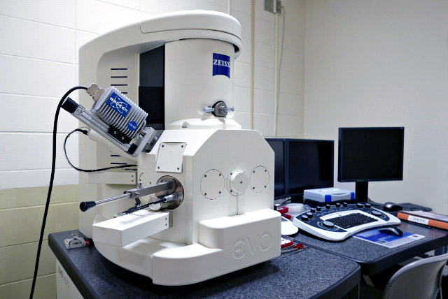
Scanning Electron Microscope with Energy Dispersive Spectrometer (SEM/EDS)
The SEM allows for high resolution images to be taken of a variety of samples by bombarding the surface of the material with electrons and detecting the secondary electron yield. A high vacuum technique, this model allows for the sampling of non-traditional materials and simplified sample preparation due to extended pressure options. The EDS allows for elemental analysis of the material imaged in the SEM.
Zeiss EVO HD with Bruker EDS
- 3.7 nm/129 eV resolution for imaging and EDS, respectively
- Extended Pressure mode
- SmartSEM and ESPRIT software
- Peltier cooling stage
- STEM capabilities
- Secondary and backscattered electron detection
- Line, point, and area mapping EDS
Contact: Sarah Lantvit
 Zeiss-EVO-HD-Bruker-EDS
Scanning Tunneling Microscope (STM)
Similar to AFM, but relies on changes in current to look at the surface, rather than changes in force.
Agilent 4400
- Imaging in atmosphere, inert gas, or solution environments
- Atomic resolution
Contact: Seth King
 Agilent-4400
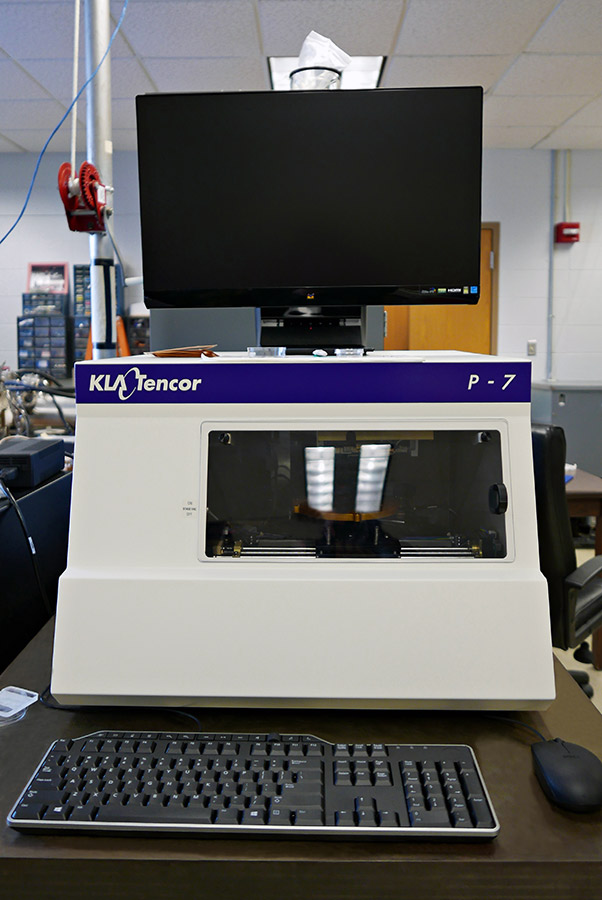
Surface Profilometer
A surface imaging technique that allows for surface mapping and roughness measurements on a larger scale than the AFM.
KLA Tencor P-7
- Stylus profiler
- 150 mm scan length standard
Contact: Seth King
 KLA-Tencor-P-7