
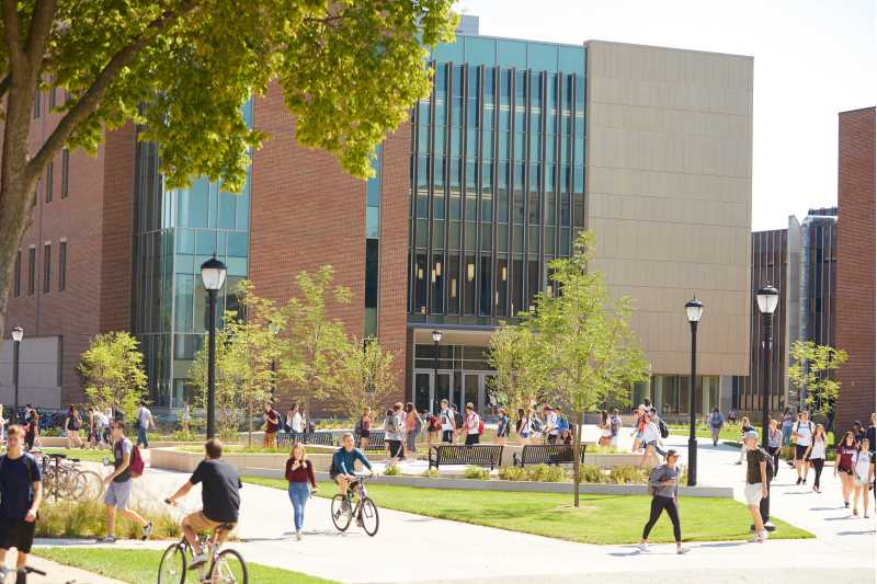
Relocation
Due to the demolition of Cowley Hall and construction of Prairie Springs Science Center Phase II, the Biology Department will be temporarily relocated to 212 Cartwright Center. We apologize for any inconvenience.
Undergraduate programs
Biology
Undergrad major Undergrad minor Teacher licenseMajor in biology! If you want to prepare for a career in healthcare, learn about organisms worldwide or help cure diseases, biology may be right for you.
Areas of study
Aquatic Science Concentration
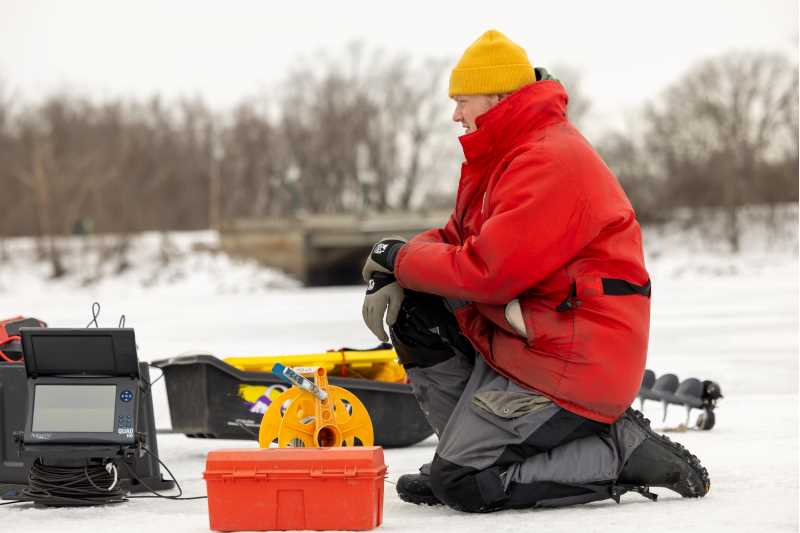
UWL offers an aquatic science concentration within the biology major to prepare students for exciting and challenging careers in the study and management of aquatic resources such as wetlands, streams, lakes, rivers, springs and groundwater.
Undergrad major View a sample plan for Aquatic Science Catalogfor Aquatic ScienceBiomedical Science Concentration
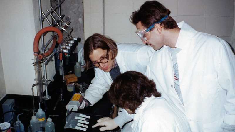
Biomedical science concentration coursework focuses on human and mammalian biology. An excellent choice for pre-med, pre-vet and pre-health professions students, the concentration includes a highly regarded, two-semester human anatomy and physiology series and additional chemistry classes.
Undergrad major View a sample plan for Biomedical Science Catalogfor Biomedical ScienceConservation Biology
Undergrad majorMolecular Genetics & Cell Biology Concentration
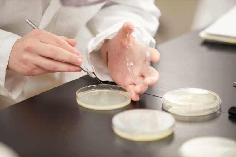
Molecular Genetics and Cellular Biology concentration coursework focuses on understanding living processes at a molecular level. Scientists are making exciting biological discoveries in these fields today whether identifying genes responsible for cancer or re-writing the genetic code of living cells.
Undergrad major View a sample plan for Molecular Genetics & Cell Biology Catalogfor Molecular Genetics & Cell BiologyPlant & Fungal Biology Concentration
Plant & Fungal Biology concentration coursework will help you learn to identify plants and fungi, study their evolution, and understand their physiology. Students can also conduct research with faculty who study plants and fungi. UWL offers one of the only plant and fungal biology degrees in the U.S.
Undergrad major View a sample plan for Plant & Fungal Biology Catalogfor Plant & Fungal BiologyScience Education
Completion of the Biology: Science Education Concentration Program and associated benchmark assessments will lead to endorsement a Middle and High School Science, grades 4-12 (2600), Wisconsin teaching license.
Undergrad major Teacher license View a sample plan for Science Education Catalogfor Science EducationZoology and Animal Physiology
Undergrad majorBiology & Physical Therapy
UWL offers an accelerated graduate program for qualified students, combining an undergraduate Biology degree with a graduate Physical Therapy degree.
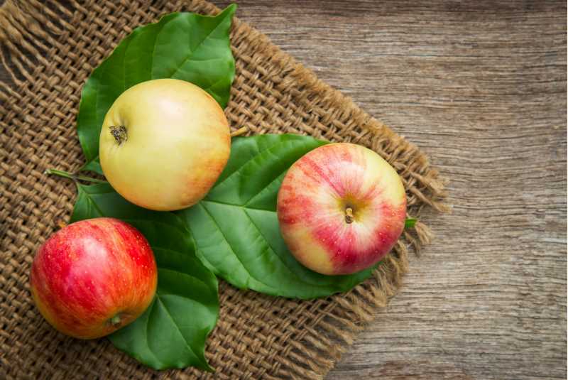
Food & Nutrition Sciences
Undergrad majorMajor in food and nutrition sciences. Gain a broad understanding and skillset related to food science, food safety, food systems and nutrition.

Nutrition
Undergrad minorWhat can you do with a nutrition minor? Broaden your knowledge and be more competitive for employment or graduate school in health and other career paths.
Related programs from other departments
Graduate programs
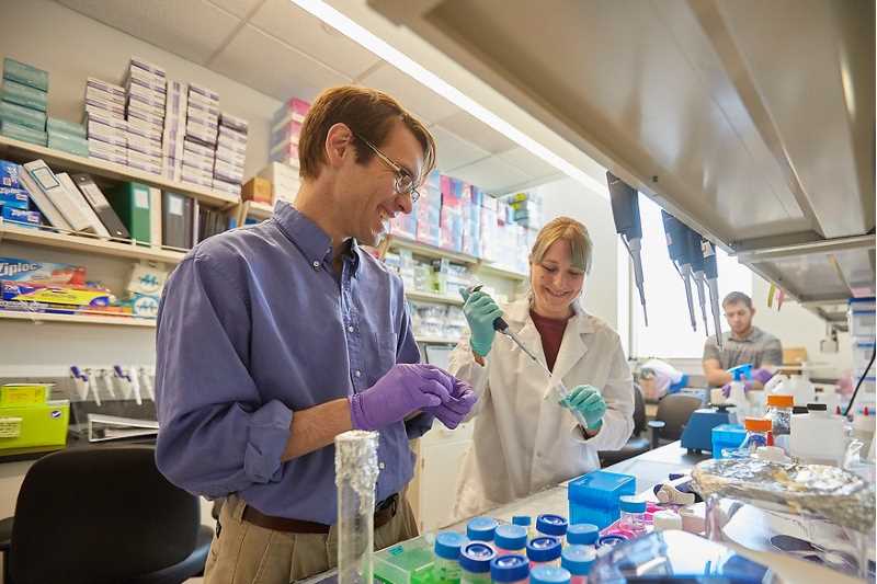
Biology
Graduate degreeAdvance your expertise in biology through hands-on research, individualized mentorship, and advanced scientific training with an M.S. in Biology.
Areas of study
Aquatic Science Concentration
Graduate degreeCellular & Molecular Biology Concentration
Graduate degreeEnvironmental Science Concentration
Graduate degreeNurse Anesthesia Concentration
Graduate degreePhysiology Concentration
Graduate degree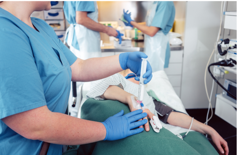
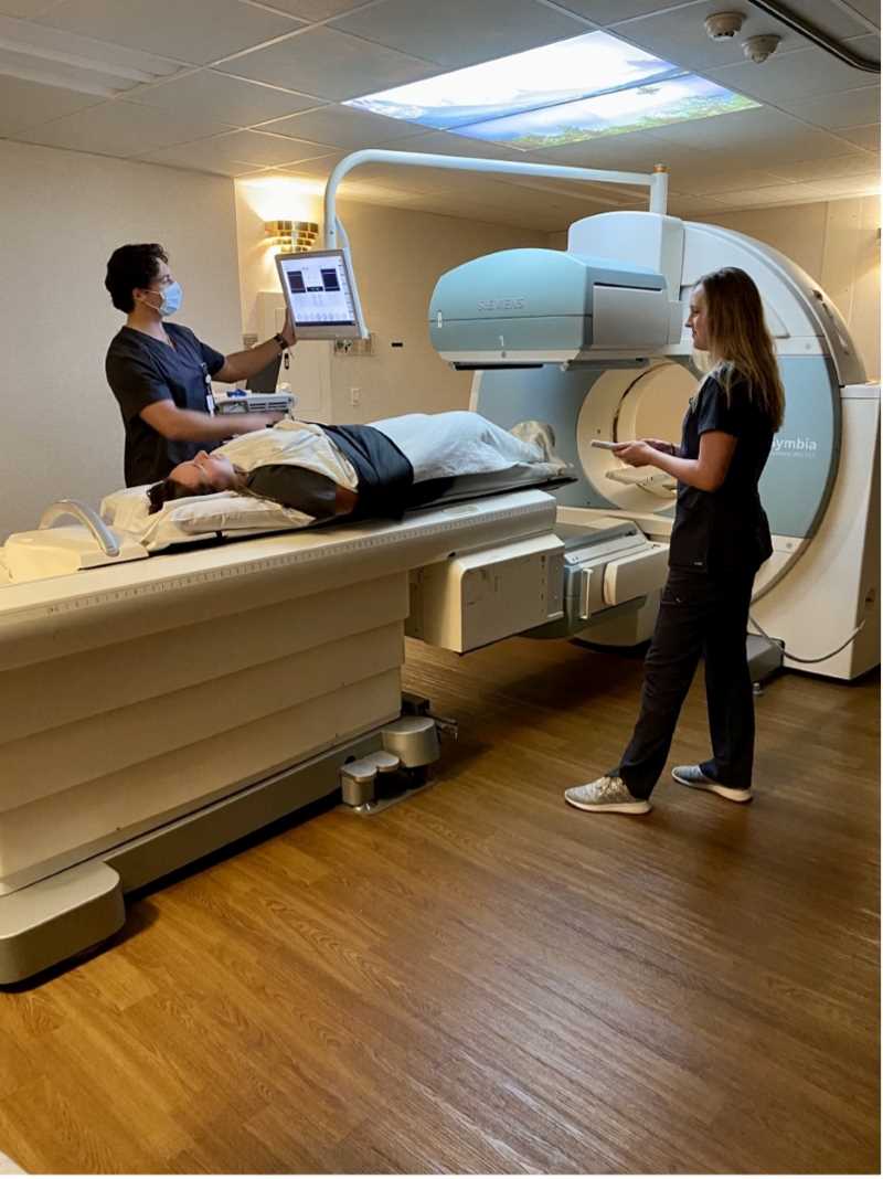
Nurse Anesthesia
Graduate degreeAdvance your nursing career — and play a vital role in patient safety, comfort and care.
Featured courses
View all coursesGet assistance
Featured event
Latest news

Upcoming Academic Calendar dates

Hear from an alum
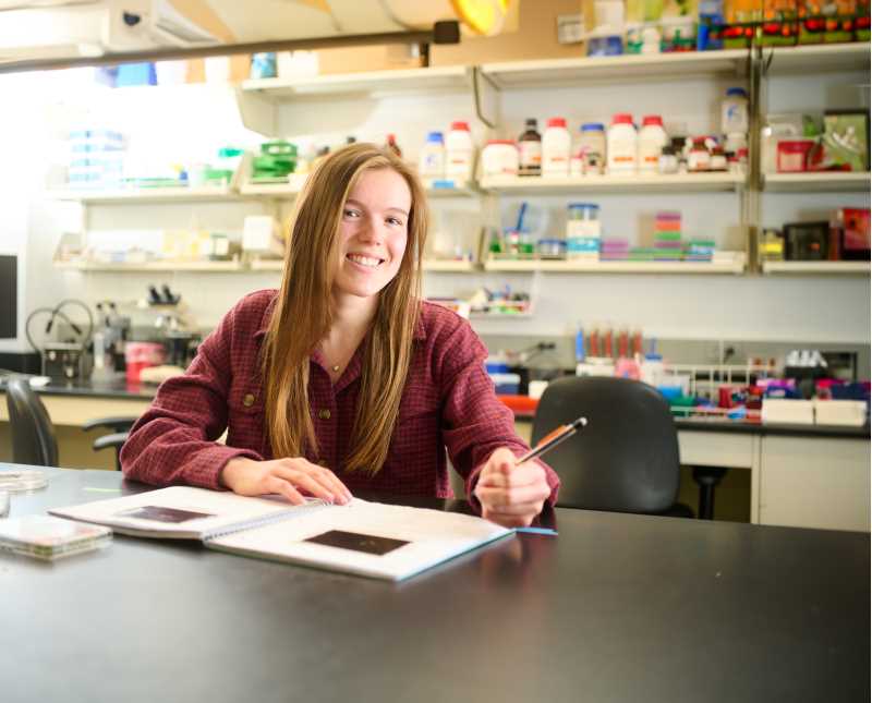
Rachel Fenske, Ph. D., RDN
The faculty in the UWL Nutrition program are engaged and knowledgeable. I had the opportunity to build my nutrition knowledge base, explore new research, and gain insight into future careers.

Hear from an alum
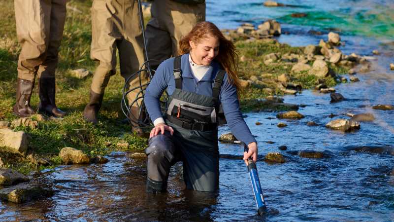
Sarah Hahn
A professor once told me, a good scientist never stops being curious. I would not be where I am today if it wasn't for the professors in the biology department constantly encouraging my curiosities.

Hear from an alum
Katherine Le Mere
The nutrition program informed me about the job possibilities in food science, and the hands-on aspect of the food science class helped me realize I would enjoy a career in the field. It also provided enough knowledge to give me the confidence to apply for a master’s degree program in food science. I can’t wait to see where this path will take me!

HIRED!

Jaime Anderson
Wisconsin Lake & Pond Resource | Aquatic Biologist
I have gained so many lifelong experiences!

Hear from an alum
Abby Siakpere
My main reasons for coming to UWL were the Biology Department, the new science labs building, and the excellent reputation the biology program has.